Deep Vein Thrombosis Treatment in Borivali
What is Deep vein thrombosis?
Deep Vein Thrombosis (DVT) refers to the formation of blood clots or thrombus within the deep veins, with the legs being the most common location. However, it can also develop in the arms or other parts of the body. In some cases, a portion of the clot, known as an embolus, can break off and travel to the lungs, resulting in a pulmonary embolism. This can block blood flow to all or a portion of the lung, leading to a medical emergency that can be life-threatening.
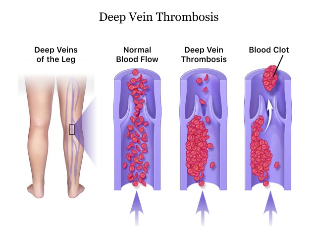
- Leg veins contain small valves that help keep blood moving toward the heart. Injury, immobility, and other factors can lead to the formation ofa blood clot
- Inside a leg vein. This is called a deep-vein thrombosis. Sometimes a piece of the clot breaks away
- (this is called an embolus) and enters the circulation. If it lodges in the
lungs, It can cause a potentially deadly pulmonary embolism.
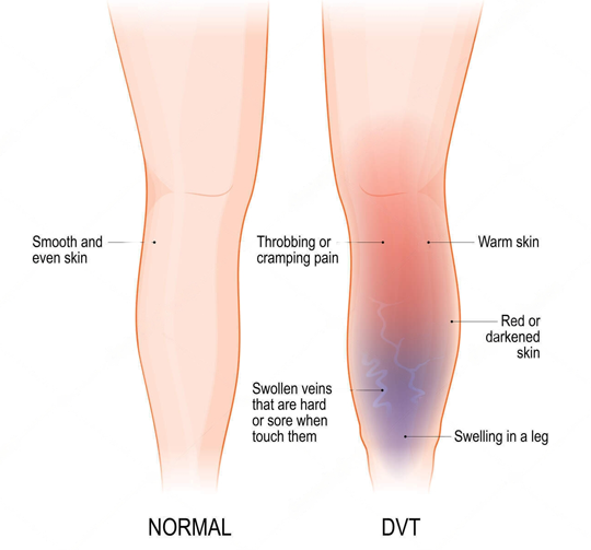
Symptoms of deep vein thrombosis
- Sudden severe swelling
- Cramp soreness in the leg
- Change in skin color on the leg
- Abdominal pain
- Chest pain and breathlessness
- Long term eczema and ulcers around ankle
What are the risk factors of DVT ?
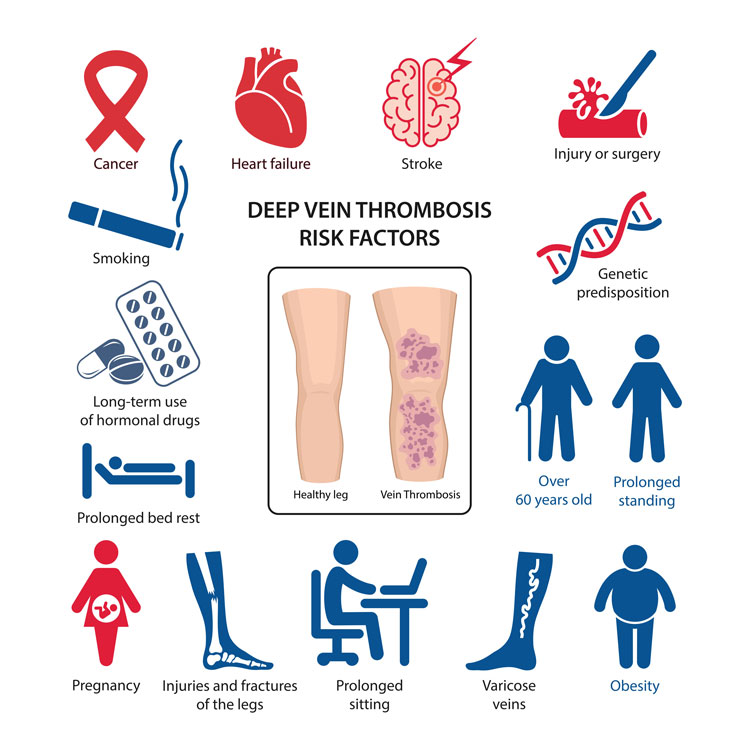
Everyone Is At Risk. Some Factors Can Increase This Risk.
HOSPITALIZATION AND SURGERY
One-half of blood clots occur during or soon after a hospital stay or surgery.
BEING IMMOBILE
Not moving for long periods of time (for example, extended bed rest or extended travel)
Other Risk Factors:
- Being older than 60s
- Pregnancy
- Birth control pills
- Being overweight
- Smoking
- Cancer
- Heart failure
- Inflammatory bowel disease
- Genetics
How to diagnose deep vein thrombosis?
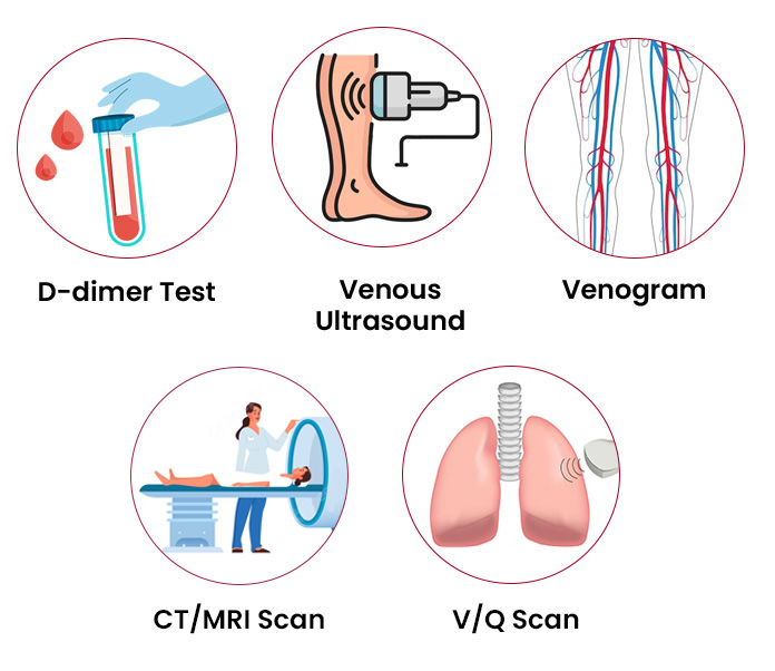
- Medical History and Physical Examination: The doctor will first discuss your symptoms, medical history, and any risk factors you may have for DVT. They will then perform a physical examination, checking for signs such as swelling, tenderness, or discoloration in the affected area.
- Ultrasound Imaging: Ultrasound is a widely used imaging technique to diagnose DVT. It uses sound waves to create images of the veins and assess blood flow. Doppler ultrasound, specifically, can help visualize blood flow and detect blood clots within the veins.
- D-Dimer Test: D-dimer is a blood test that measures the presence of a substance produced when a blood clot dissolves. Elevated levels of D-dimer may indicate the presence of a blood clot, but it is not conclusive for diagnosing DVT. This test is often used in conjunction with other diagnostic methods.
- Venography: Venography involves injecting a contrast dye into the veins and taking X-ray images. This test helps visualize the veins and any potential blockages caused by blood clots.
- Magnetic Resonance Imaging (MRI): In certain cases, an MRI scan may be performed to obtain detailed images of the veins and identify any blood clots present.
Diagnosis of Pulmonary Embolism
- CT Venography: Creates 3D images that finds detects pulmonary embolism within the arteries of the lungs.Outlines the pulmonary arteries with the use of contrast.
- Pulmonary angiography: Provides a clear picture of the blood flow in the arteries of the lungs. It’s the most accurate way to diagnose a pulmonary embolism.
- Ventilation perfusion Scan: Here a small amount of a radioactive substance called a tracer is injected into a vein in your arm. The tracer maps blood flow, called perfusion, and compares it with the airflow to your lungs, called ventilation.
What factors determine Treatment for Deep Vein Thrombosis?
Specific treatment will be determined by our expert Vascular Interventional Radiologist based on how long the clots have been present and various other factors.
- How old you are
- Your overall health and medical history
- How sick you are
- The location of the clot
- How well you can handle specific medicines, procedures, or therapies
- How long the condition is expected to last
- Your opinion or preference
The goal of treatments is to prevent the clot from getting larger, to prevent a blood clot from traveling to the lungs (PE), to decrease the chance of another blood clot forming, and to prevent the development of Post Thrombotic Syndrome (PTS).
How do Interventional radiologist treat deep vein thrombosis?
Based on how long the clots have been present, DVT is categorized into two types: Acute and chronic.
Acute DVT
• Medication ( Blood thinners)
The standard of care for the treatment of acute DVT is blood thinning medication (anticoagulation). Blood thinning medications work by allowing blood to flow around a trapped clot while at the same time preventing the clot from traveling to the lungs.
However, these medications do not remove the existing clot from the vein. Blood thinning medications rely on the body’s natural defenses to “dissolve” the clot which can take several months and often results in the vein remaining permanently blocked.
Therefore, our trained Interventional Radiologist offers the latest interventional radiology treatment methods to prevent these complications, individualized and personalized based on your current condition.
For Deep Vein Thrombosis Treatment in Borivali, Call on 90040 93090
• IVC filter
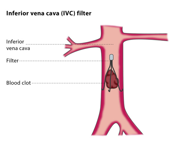
If blood-thinning medications are not recommended or there is a large clot burden, a small filter may be implanted in the inferior vena cava (IVC), the large vein that returns blood back to the heart.
This filter, called an IVC filter, prevents the clot from traveling to the lungs and heart by trapping it while allowing normal blood to flow around it.
• DVT Thrombolysis (Clot Busters)
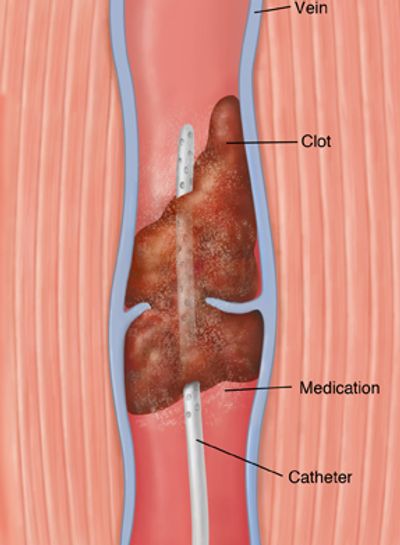
For patients with symptomatic acute DVT and large clots, a minimally-invasive procedure known as DVT thrombolysis is commonly performed to rapidly reduce symptoms and remove the clot from the deep veins.
DVT thrombolysis involves inserting a small catheter into the leg using ultrasound and X-ray guidance. Clot-dissolving medications are used to dissolve the clot.
While there is a small risk of bleeding from the procedure, DVT thrombolysis can speed the recovery from DVT and help to minimize future scarring of the veins from DVT. This potentially reduces the chances of developing a severe condition called Post Thrombotic Syndrome (PTS).
DVT thrombolysis is done on an inpatient basis with about a two-night hospital stay depending on the age of the clot. Patients with the best outcomes are those who have had symptoms for 14 days or less as this is when the clot is “fresh”. Those with symptoms ranging from 10-30 days also do well.
• Thrombectomy (Clot Suction)
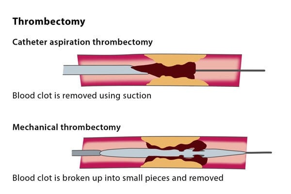
When clot busters drugs cannot be administered or many a time in combination with Thrombolysis, a newer method for treatment can be offered to you which is mechanical thrombectomy.
Here small catheters and devices are introduced into the blocked veins and the clots are broken into small pieces and then aspirated out of the vein (clot suction) to restore normal blood flow.
Chronic DVT and Post Thrombotic Syndrome (PTS)
For patients who have had clots for months to years, or who continue to experience symptoms after medication or other therapies, and those who have developed post-thrombotic syndrome due to chronic DVT, we offer advanced treatment options to relieve their symptoms and improve their quality of life.
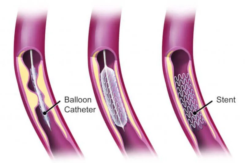
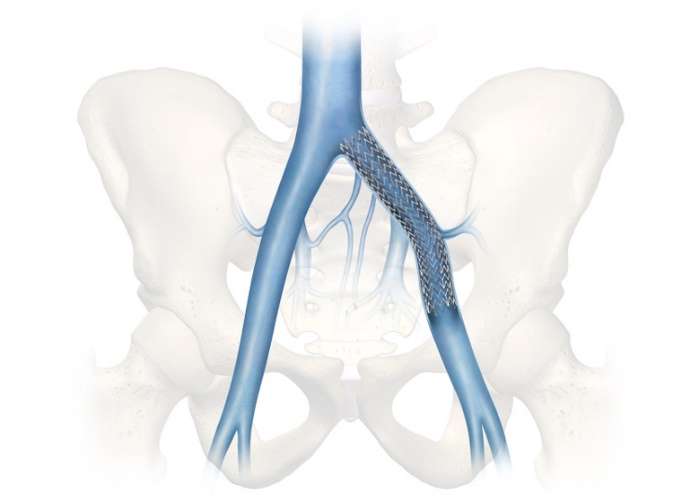
- Angioplasty and Stenting: With an angiogram one or more areas of narrowing of the vein that caused the blood clot to form can be determined. We can then treat this with angioplasty (widening the vein with a balloon), or by inserting a mesh-like tube called a stent to keep the vein open.
What are the preventive measures for deep vein thrombosis?
- Engage in regular exercise to promote healthy blood circulation.
- Quit smoking to improve vascular health and decrease the likelihood of clot formation.
- Maintain a healthy weight and BMI to alleviate strain on veins.
- Avoid prolonged sitting by taking regular breaks and moving your feet to prevent blood pooling.
- Adhere to prescribed medications as directed by your doctor to manage clotting risk.
- Keep blood pressure under control to prevent damage to blood vessels.
- Inform your doctor about any family history of DVT to assess potential risk factors.
- Wear compression stockings to minimize swelling and reduce the risk of clot development.
What are the complications of DVT?
- Pulmonary Embolism: It happens when part of the clot, called an embolus, separates from the vein. It goes to the lungs and cuts off blood flow. A PE may develop quickly. It’s a medical emergency and may cause death.
- Chronic venous insufficiency: This may happen following DVT of a leg vein. It means that a vein no longer works as well. It’s a long-term condition where blood stays in the vein instead of flowing back to the heart. Pain and swelling in the leg are common symptoms.
- Post-thrombotic syndrome: This may also happen after DVT of a leg vein. It’s a long-term problem with pain, swelling, and redness. Ulcers and sores can also happen if the condition isn’t treated early. These complications and related symptoms may make it hard to walk and take part in daily activities.
Meet Our Interventional Radiologist

Dr. Kunal Arora
M.B.B.S., M.D. Radiodiagnosis, F.V.I.R
VASCULAR AND INTERVENTIONAL RADIOLOGIST
Dr. Kunal Arora is a Vascular and Interventional Radiologist with over 5 years of experience in this field. Practicing at Endovascular Care Center in Mumbai, He specializes in treating various diseases in a minimally invasive way without the need for surgery.
His areas of interest include the management of varicose veins, DVT, peripheral arterial disease, embolization, oncologic intervention, hepatobiliary interventions, and dialysis AV fistula salvage.

